Health
Evidence for SARS-CoV-2 coronavirus particles in heart tissue
Researchers in Milan, Italy, found that severe acute respiratory syndrome coronavirus 2 (SARS-CoV-2) was found in the heart tissue of patients with coronavirus disease 2019 (COVID-19) who died without signs of respiratory failure. Identified the presence of heart involvement.
The virus is present in cardiomyocytes of all 6 patients and active viral transcription of RNA for the virus Spike protein Had occurred. Spike proteins are surface structures used by SARS-CoV-2 to bind and access host cells.
Damage to cardiomyocytes ranged from lack of cell death and no intracellular changes to structural abnormalities and intracellular edema.
Gaetano Pietro Brufamante (University of Milan) and colleagues say the findings show that COVID-19 survivors need to be revised to monitor guidelines once they are discharged from the hospital.
Long-term cardiovascular outcomes may be similar to those seen in survivors of the SARS-CoV-1 outbreak from 2002 to 2003, with 40% of individuals recovering during the 12-year follow-up period. The team warns that they have developed cardiovascular abnormalities.
A preprinted version of the paper is available on the server medRxiv* Articles undergo peer review.
COVID-19 can contain the cardiovascular system
As the SARS-CoV-2 pandemic continues to dominate the world, the virus continues to infect hundreds of thousands of people every day and COVID-19 mortality rates continue to increase worldwide.
In addition to virus-induced respiratory disease, research has shown that it may be involved in several other organs, including the cardiovascular system.
Twenty to forty percent of hospitalized patients experience heart symptoms ranging from chest pain to arrhythmias and cardiogenic shock.
In addition, up to 7% of COVID-related deaths are associated with myocarditis, said Brufamante and the team.
However, so far it is not clear whether cardiovascular involvement is due to cells directly damaged by the virus or secondary to the involvement of hyperreactive immune responses. Hypoxia due to acute respiratory distress syndrome (ARDS) can also cause myocardial damage.
On the other hand, SARS-CoV-2 has been confirmed in vascular endothelial cells of patients and cardiac macrophages of patients with myocarditis. In addition, viral RNA has previously been identified throughout heart tissue, regardless of heart phenotype, the researchers say.
“Nevertheless, immunohistochemistry for the detection of viral antigens in cardiomyocytes has never been demonstrated,” the team wrote.

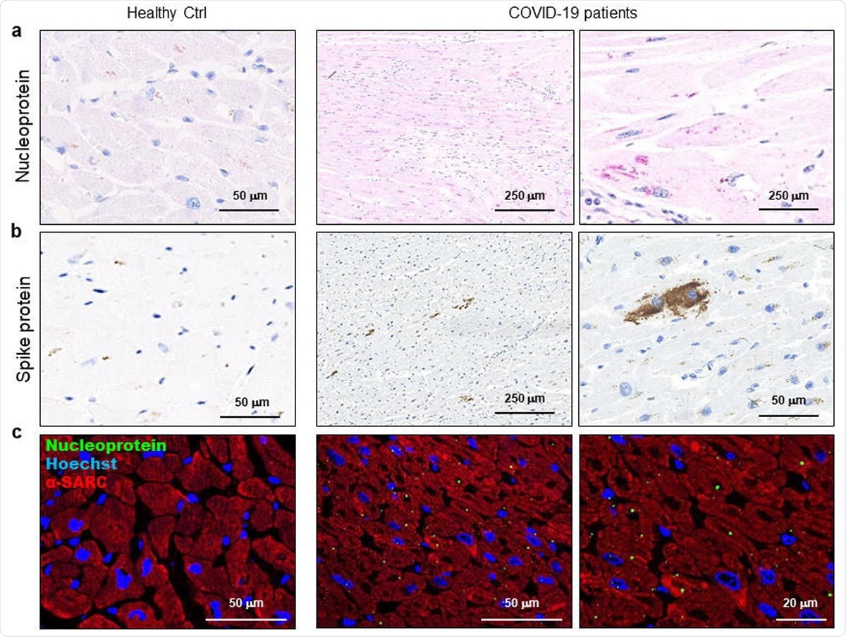
SARS-CoV-2 protein can be detected in cardiomyocytes of COVID-19 patients. ab) Representative of immunohistochemical assay of 3 μm slides of formalin-fixed paraffin-embedded left ventricle specimens from healthy controls (Healthy Ctrl, left panel) and COVID-19 patients (COVID-19, middle and right panels) for SARS-CoV. Image-2 nuclear protein (a, red) and spike protein (b, brown). c) Representative of a 3 μm slide immunofluorescence assay of formalin-fixed paraffin-embedded left ventricular specimens from healthy controls (Healthy Ctrl, left panel) and COVID-19 patients (COVID-19, middle and right panels) for SARS-CoV. Image-2 nuclear protein (green) and sarcomeric α-actin (α-SARC, red). Nuclei are stained with Hoechst (blue). Magnification as a scale bar.
What was included in the study?
The researchers tested the presence of SARS-CoV-2 in the heart tissue of six COVID-19 patients (54 to 69 years old) who died of respiratory failure without showing signs of heart failure.
Using chip-based digital PCR, the team detected SARS-CoV-2 viral RNA in cardiomyocytes from heart tissue samples taken from all six patients. Virus spike proteins in cardiomyocytes were also identified using Western blot, immunohistochemistry, and immunofluorescence, and electron microscopy revealed the presence of intact virus particles.
Interestingly, the researchers found that spike protein expression varied between patients and hypothesized that the viral genome was actively transcribed in cardiomyocytes.
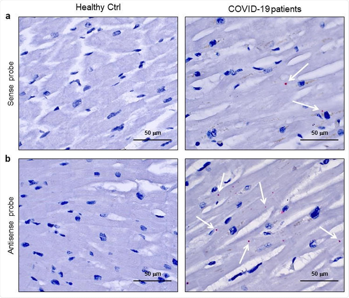
To test the presence of SARS-CoV-2 as a transcriptionally active virus in cells, the team performed an RNAScope assay using two probes that recognize spike protein sense and antisense RNA, I made it possible to distinguish the transcript. Signals were obtained with both probes in all patient samples, but the most abundant staining was observed with probes that detected actively transcribed virus.
The researchers say this is the first study to identify SARS-CoV-2 RNA localization in cardiomyocytes and active viral transcription of both sense and antisense RNAs of the viral spike proteins.

SARS-CoV-2 sense and antisense RNAs are localized in cardiomyocytes of COVID-19 patients. ab) Representative of spatially resolved viral RNA detection by RNAScope assay at 3 μm slide in formalin-fixed paraffin-embedded left ventricle preparations from healthy controls (Healthy Ctrl, left panel) and COVID-19 patients (COVID-19, middle and right panels). Images for SARS-CoV-2 sense (a) and antisense (b) probes for spike protein RNA sequencing. Fast Red dots indicate the presence of viral RNA (white arrow). Nuclei are counterstained with hematoxylin. Magnification as a scale bar.
“Premature, asymptomatic signs of cell damage”
The patterns of cardiomyocyte injury varied. Virus-containing cells did not appear to undergo apoptosis, but showed intracellular changes such as increased intracellular volume and intracellular edema.
Some cardiomyocytes, including SARS-CoV-2, appeared normal, whereas other myocytes showed excessive intracellular regions between the sarcolemma and sarcomeric structures, explaining the observed intracellular edema. The authors say it may be.
“Some cardiomyocytes show altered cell structure with increased cell area and intracellular edema, suggesting cardiomyocyte swelling,” the team wrote. “This microscopic event could be interpreted as an asymptomatic sign of premature cell damage, leading to the formation of myocardial interstitial edema.”
Post-Discharge Monitoring Guidelines Need Revision
Researchers say that heart tissue containing contractile cell compartments can be infected with SARS-CoV-2, which initially causes microscopic cardiomyocyte changes that do not lead to clinically relevant macroscopic heart damage Researchers say research has shown that
“Our key study specifically suggests the need to redefine post-discharge monitoring guidelines to survive follow-up in COVID-19 patients without a pronounced cardiac phenotype.” Brufamante and colleagues say.
“The long-term cardiovascular outcomes of patients with COVID-19 may reflect those with SARS who developed cardiovascular abnormalities in 40% of those who recovered at a long-term follow-up of 12 years,” they conclude. I will.
*Important Notices
medRxiv It publishes preliminary, non-peer reviewed scientific reports and should not be considered conclusive and should not guide clinical practice/health-related behavior or be treated as established information.
..
What Are The Main Benefits Of Comparing Car Insurance Quotes Online
LOS ANGELES, CA / ACCESSWIRE / June 24, 2020, / Compare-autoinsurance.Org has launched a new blog post that presents the main benefits of comparing multiple car insurance quotes. For more info and free online quotes, please visit https://compare-autoinsurance.Org/the-advantages-of-comparing-prices-with-car-insurance-quotes-online/ The modern society has numerous technological advantages. One important advantage is the speed at which information is sent and received. With the help of the internet, the shopping habits of many persons have drastically changed. The car insurance industry hasn't remained untouched by these changes. On the internet, drivers can compare insurance prices and find out which sellers have the best offers. View photos The advantages of comparing online car insurance quotes are the following: Online quotes can be obtained from anywhere and at any time. Unlike physical insurance agencies, websites don't have a specific schedule and they are available at any time. Drivers that have busy working schedules, can compare quotes from anywhere and at any time, even at midnight. Multiple choices. Almost all insurance providers, no matter if they are well-known brands or just local insurers, have an online presence. Online quotes will allow policyholders the chance to discover multiple insurance companies and check their prices. Drivers are no longer required to get quotes from just a few known insurance companies. Also, local and regional insurers can provide lower insurance rates for the same services. Accurate insurance estimates. Online quotes can only be accurate if the customers provide accurate and real info about their car models and driving history. Lying about past driving incidents can make the price estimates to be lower, but when dealing with an insurance company lying to them is useless. Usually, insurance companies will do research about a potential customer before granting him coverage. Online quotes can be sorted easily. Although drivers are recommended to not choose a policy just based on its price, drivers can easily sort quotes by insurance price. Using brokerage websites will allow drivers to get quotes from multiple insurers, thus making the comparison faster and easier. For additional info, money-saving tips, and free car insurance quotes, visit https://compare-autoinsurance.Org/ Compare-autoinsurance.Org is an online provider of life, home, health, and auto insurance quotes. This website is unique because it does not simply stick to one kind of insurance provider, but brings the clients the best deals from many different online insurance carriers. In this way, clients have access to offers from multiple carriers all in one place: this website. On this site, customers have access to quotes for insurance plans from various agencies, such as local or nationwide agencies, brand names insurance companies, etc. "Online quotes can easily help drivers obtain better car insurance deals. All they have to do is to complete an online form with accurate and real info, then compare prices", said Russell Rabichev, Marketing Director of Internet Marketing Company. CONTACT: Company Name: Internet Marketing CompanyPerson for contact Name: Gurgu CPhone Number: (818) 359-3898Email: [email protected]: https://compare-autoinsurance.Org/ SOURCE: Compare-autoinsurance.Org View source version on accesswire.Com:https://www.Accesswire.Com/595055/What-Are-The-Main-Benefits-Of-Comparing-Car-Insurance-Quotes-Online View photos
Pictures Credit
to request, modification Contact us at Here or [email protected]


